X-Rays, CAT scans and Medical Images
Activities
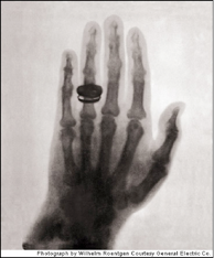
Photograph by Wilhelm Roentgen: Berta Roentgen’s hand with wedding band. Courtesy General Electric Co.
In 1895, Wilhelm Conrad Röntgen created the first plates of X-ray photographs, taking shadowy images of his wife’s hand. Similar to the images we saw projected in the CAVE activities, this was a profound medical advance as for the first time it allowed us to see internal body structures non-invasively. Röntgen’s discovery earned him very first Nobel Prize (Physics, 1901), but contributed to his death in 1923 from Leukemia due to X-ray exposure.
As important as X-Rays are, they offer only a single planar image of even the most complex anatomical structures. I.e. they are like single clues in the Flatland or CAVE games. Extending imaging techniques to capture 3-dimensional structures in their entirety via tools like CAT scans, MRI, and PET scans – accomplished over the last 35 years or so – has revolutionized modern medicine by allowing us to visualize every bodily organ with tremendous resolution as a real 3-dimensional object.
CAT scans – like the other imaging technology – work by little more than a combination of the slicing and projection techniques we’ve considered here. Using a circular array of X-Rays and detectors, numerous images from different perspectives along an axial slice (the “A” in CAT) are relayed to a computer (the “C” in CAT). Using mathematical algorithms known as tomographic reconstruction (the “T” in CAT), the computer obtains a high-resolution image of an axial slice. This process is repeated to obtain hundreds of slices and the computer integrates these much like we did in constructing sliceforms to provide a true 3-dimensional map of the solid being analyzed. The solid can be seen using interactive computer graphics or even through stereolithographic models like those that are made for hip replacement surgery. Those who make this possible are Flatland game experts – with real life rewards. For accessible further information, see Sochurek, 1988 and the Project 2000 Internet site.
CAT Activities:
-
1. Working in small groups, draw axial cross sections of different regions of your bodies. (This is the view one would see as if they were passing through Flatland standing up.) Check your accuracy using one of the many available Visible Human Program viewers available online (e.g. the NPAC/OLDA Visible Human Viewer at www.dhpc.adelaide.edu.au/projects/vishuman2/VisibleHuman.html ; for a full list of viewers, consult the U.S. National Library of Medicine at http://www.nlm.nih.gov/research/visible/applications.html .)
2. Working in small groups, play the Flatland game using human body parts as the mystery objects.
3. Working in small groups, play the Flatland game using a variety of different animals.
Copyright 2011 Julian Fleron and Volker Ecke.